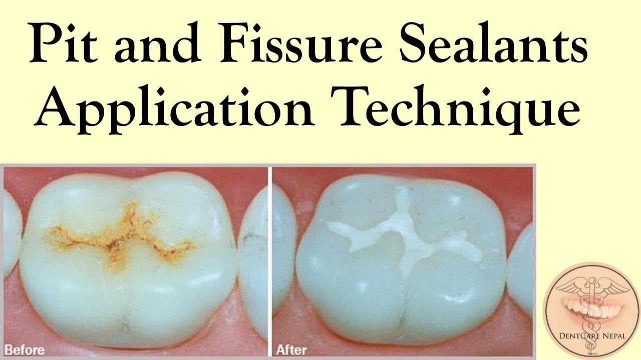
The term pit and fissure sealant (PFS) is used to describe
a material, i.e., introduced into the occlusal pits and fissures of caries-susceptible teeth, thus forming a micromechanically bonded, protective layer cutting access of
caries producing bacteria from their source of nutrients.1
Buonocore’s classic study of 1955 marked the start
of a major revolution in the clinical practice of dentistry.
The first clinical benefit from Buonocore’s work was the
introduction of the first dental PFS, Nuva-Seal (LD Caulk)
in February 1971, along with its curing initiator, and
ultraviolet light source, the Caulk Nuva Lite
it took several more years before the sealant technique,
and other clinical innovations that have resulted from
Buonocore’s work, began to be adopted in clinical dentistry to any significant degree. Still, more than 30 years
after the introduction of PFS to the dental market place,
the profession has not embraced the procedure to the
extent that available scientific data would expect.2
While levels of tooth decay (dental caries) in children
and adolescents have declined in many parts of the world
in recent decades, caries remains a public health problem
in many countries.
CLINICAL INDICATIONS FOR PFS
Indications
• The occlusal surfaces of permanent teeth having welldefined pit and fissures and/or deep fossae. Occasionally, primary molars with significantly deep grooves
or pits may be sealed
• Stained or slightly white pit and fissure, especially in
patients with high caries incidence
• Buccal and lingual grooves when only the appropriate
teeth have erupted sufficiently to be free of gingival
and operculuctum contact
• Incisors with lingual pits.
Contraindications
• Synthetic porcelain restorations
• Veneers
• Amalgam restorations
• Gold foil restorations, inlays, onlays, or crown
• Evidence of caries on occlusal or interproximal
surfaces
• Teeth that cannot be sufficiently isolated
• Sealing margins of existing nonresin restorations
• Vital dentin, which is more sensitive than enamel and
has a much poorer retention rate
• In children who are too young to cooperate during
the procedure.
Follow-up and Review
All sealed surfaces should be regularly monitored clinically and radiographically. Bitewing radiographs should
be taken at a frequency consistent with the patient’s risk
status, especially where there has been doubt as to the
caries status of the surface prior to sealant placement.
The exact intervals between radiographic reviews will
depend not only on risk factors, which may change over
time, but also on monitoring of other susceptible sites,
e.g., proximal surfaces.20
RECOMMENDATIONS
• Apply sealants to the permanent molar teeth of children and youth who are identified at risk by a caries
risk assessment.
• Place sealants on teeth as soon as possible after eruption; however, the length of time after eruption should
not be a barrier to placement of sealants.
• Use resin-based sealant materials to seal teeth, for
increased retention of dental sealants.
• Disseminate the review findings to peel dentists,
dental hygienists, level II dental assistants, and other
health care providers to promote the use of PFS.
CONCLUSION
Most of the carious lesions that occur in the mouth occur
on the occlusal surfaces. Which teeth will become carious
cannot be predicted; however, if the surface is sealed with
a pit and fissure sealant, no caries will develop as long as
the sealant remains in place. Sealants are easy to apply,
but the application of sealants is an extremely sensitive
technique. So only trained staff should be allowed to
place the sealants


