“Definitions, Types & Management”
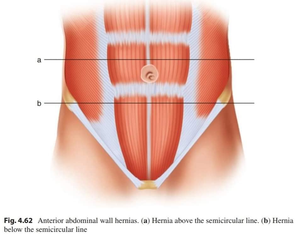
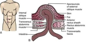
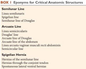
The first is the arcuate line, which is a transverse line that marks the point where the posterior surface of the rectus abdominus muscle is only covered by transversalis
fascia and peritoneum and is the site of entry of the inferior epigastric vessels into the rectus muscle. Above arcuate line,rectus abdominus is surrounded by an anterior layer of the rectus sheath and a posterior layer.
This can also be confusing because of varying definitions in the literature, such as a line marking the transition from transversus abdominis muscle
to its aponeurosis, from the costal margin to pubic tubercle versus the site of splitting of internal oblique aponeurosis at the lateral edge of rectus abdominis muscle.
(arcuate line, fold, or line of Douglas) marks the caudal end of the posterior lamina of the aponeurotic rectus sheath, below the umbilicus and above the pubis. Unfortunately, the semilunar and semicircular lines are not easily seen in the operating room.
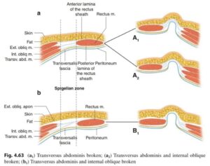
It is a small strip of aponeurosis of the transversus abdominis muscle bounded laterally by linea semilunaris and medially by the lateral margin of the rectus abdominis muscle.
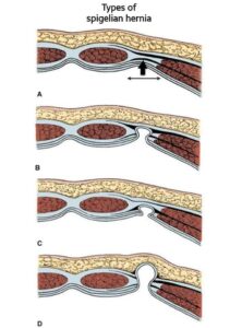
Spigelian hernias usually
are located at the junction of the arcuate line and the semilunar line in the Spigelian fascia. However, the exact location of the arcuate line is variable, which gives rise to the concept of the “Spigelian hernia belt,” which is located between horizontal lines drawn just below the umbilicus cranially and at the anterior superior iliac spines caudally and is usually about 6 cm in length. Spigelian hernias can occur anywhere along the Semilunar line, but 90% occur in the Spigelian hernia belt.
?????????:
•Spigelian hernias should be repaired due to their higher risk of incarceration and vague physical diagnosis. Surgical repair can be performed via open or minimally invasive techniques.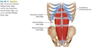

•There is little data on the benefits of prosthetic mesh in these hernias due to their rarity. However, for moderately sized defects (>2 cm) we recommend prosthetic reinforcement in open and minimally invasive cases.
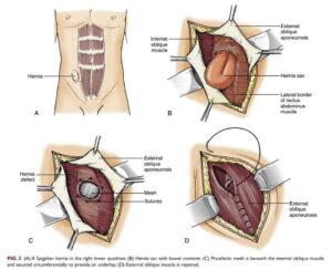
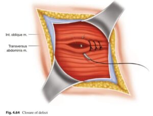
•???? repair involves a direct cutdown (typically transverse) overlying the hernia and the opening of the external oblique fascia to expose the posterior wall defect. The surgeon can then utilize a subway technique or place the mesh anterior to the posterior fascia following primary closure. The external fascia is then closed primarily.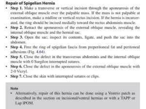


Hernia repair is most commonly performed transabdominally but may also be attempted extraperitoneally by accessing the space of Retzius. If approaching the repair from the peritoneal cavity, an ???? may be placed. Alternatively, a peritoneal flap can be created with the development of the preperitoneal space & reduction of the hernia sac before the mesh insertion. If placing mesh in the preperitoneal space, a coated, or composite, mesh is not necessary.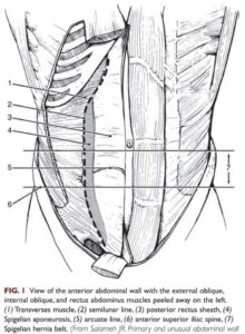

???; ☆Current surgical therapy 13th ed, 2020.
☆ Surgical Anatomy and technique 5th ed, 2021.
☆ Maingot’s Abdominal Operations, 13th ed.
☆The Art of Hernia Surgery, 2018.
