back to top
Thursday, July 25, 2024
Home Blog

Oncotype Dx – Predicts how likely6 cancer comes back or not.

Oncotype DX

1. What is the Oncotype DX test?
2. Who is Oncotype DX for?
3. Why is it used? 
4. How does it work?
5. Oncotype DX score
6. Availability and cost
7. Oncotype DX Breast DCIS Score test

1. What is the Oncotype DX test?

Oncotype DX is a test that predicts how likely breast cancer is to spread to somewhere else in the body (secondary breast cancer) within 10 years of diagnosis, in people who will be taking hormone therapy for at least five years.

2. Who is Oncotype DX for?

The test is suitable for people recently diagnosed with early stage invasive breast cancer that:

  • Has not spread to the lymph nodes under the arm
  • Is oestrogen receptor positive (ER+)
  • Is HER2 negative (HER2-)

Sometimes the test may be considered for people whose breast cancer affects one to three lymph nodes under the arm. 

The test is not suitable for people whose breast cancer is oestrogen receptor negative or HER2 positive. 

3. Why is it used?

Your specialist may recommend the Oncotype DX test if the benefit of chemotherapy for you is less clear.

Chemotherapy may be given after surgery to reduce the risk of breast cancer coming back in future.

Whether you’re offered chemotherapy depends on a number of features of your breast cancer. These include:

  • The size and grade of the cancer
  • Whether it has spread to any of the lymph nodes under the arm
  • Whether the cancer is hormone receptor and HER2 positive or negative

For some people the benefit of chemotherapy is clear, but for others it’s less clear.

4. How does it work?

The test is done on a small amount of breast cancer tissue already removed (for example during a core biopsy or surgery).

The tissue is sent to a laboratory in the USA, where the test is carried out. The test looks at groups of genes found in breast cancer.

The results are given separately from your pathology report and are sent to your specialist within 10 to 14 days. 

If your specialist has recommended you have hormone therapy before surgery, the test must be done on the tissue removed by a core biopsy, before you start hormone therapy.
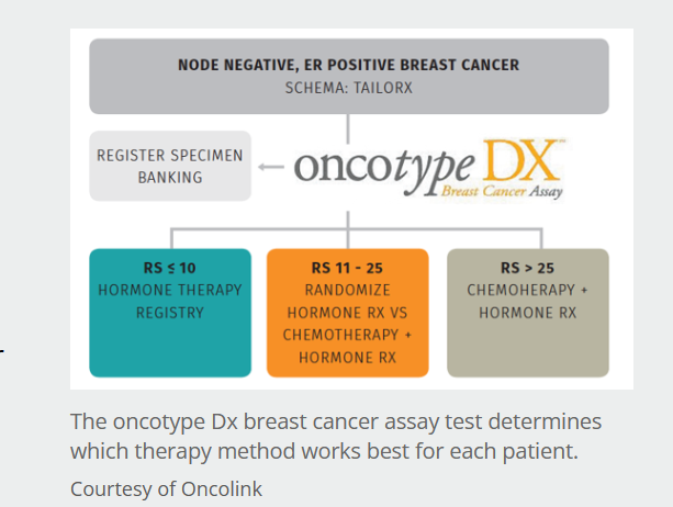
Oncotype Dx - Predicts how likely6 cancer comes back or not.

5. Oncotype DX score

The test gives a score, known as a Recurrence Score, from 0 to 100. 

The higher the score, the more likely breast cancer is to come back and the more likely you are to benefit from having chemotherapy as well as hormone therapy. 

Your specialist will use the score, along with other information about your breast cancer, to help decide whether chemotherapy would benefit you. 

Women over the age of 50

For women over 50:

  • A score of 0–25 means you’re unlikely to benefit from having chemotherapy in addition to hormone therapy
  • A score of 26 or above means you’re likely to benefit from having chemotherapy in addition to hormone therapy

Women aged 50 or younger

For women aged 50 or under:

  • A score of 0–15 means you’re unlikely to benefit from having chemotherapy in addition to hormone therapy
  • A score of 16 or above means your specialist will discuss your test result with you to help decide if you’re likely to benefit from chemotherapy

6. Availability and cost

If you don’t have any lymph nodes that contain cancer

If you don’t have any positive lymph nodes, Oncotype DX is available on the NHS across the UK.

If you have between 1 and 3 lymph nodes that contain cancer

If you have between 1 and 3 positive lymph nodes, you may sometimes be able to have the Oncotype DX test on the NHS. Check with your treatment team. 

Most private healthcare companies will also cover the cost of the test. 

You can also pay for the test yourself, but your treatment team will need to order the test for you and will be sent the results to discuss with you. It costs around £3,000. 

7. Oncotype DX Breast DCIS Score test

There’s a specific Oncotype DX test for people with ductal carcinoma in situ (DCIS) who have had breast-conserving surgery (also known as wide local excision or lumpectomy).

Oncotype DX for DCIS can predict the risk of the cancer coming back after surgery. The results of this test may help you and your treatment team decide whether you’re likely to benefit from radiotherapy.

It’s not routinely used on the NHS as there’s less evidence of its benefits for people with DCIS. Some private healthcare providers may cover the cost and you can also pay for the test yourself. For more information, talk to your treatment team.  

Scientists discover ‘critical’ link between autism and a virus-updates

A virus common in newborn babies may increase their risk of developing autism, a study suggests. 

By examining medical records of nearly 3million infants, researchers found those born with cytomegalovirus (CMV) were two-and-a-half times more likely to be ..This seemingly harmless cold-like bug affects roughly one in every 200 babies, and is the most common infection present at birth in the US.

Yet doctors in the US don’t currently screen for the common virus, which is passed from pregnant women to their fetus in the womb.

for details click link below in details

MSN health

Read more

Diffuse Dermal Angiomatosis of the Breast

Diffuse dermal angiomatosis is rare and usually considered a variant of reactive angioendotheliomatosis. It generally involves the extremities of patients with severe vascular disease and other comorbidities. Two patients with breast involvement have been described; however, neither had a relevant medical history or a vaso-occlusive disorder, but both had large pendulous breasts, and 1 was positive for IgM anticardiolipin and antinuclear antibodies.

Diffuse dermal angiomatosis (DDA) is a benign, cutaneous vascular disorder. It was first described as a subtype of reactive angioendotheliomatosis (RAE) in 1994 by Krell et al. More recently, DDA has been recognized as a distinct, rare variant of RAE associated with smoking, trauma, underlying vaso-occlusion, and hypercoagulability. Clinically, DDA manifests with tender, violaceous-to-erythematous ulcerated plaques and purpuric papules., Lesions may also have central ulceration with surrounding tissue necrosis. Lesions tend to persist and progressively enlarge. While the extremities are the most commonly reported location, there have been several reported cases of DDA of the breast (DDAB), indicating that DDAB is likely more prevalent than previously thought., Herein, we report a case of bilateral DDAB and review the management strategies utilized for this disease to date.

DDA is a unique variant of RAE commonly reported on the lower extremities and, more recently, in women with large, pendulous breasts., DDAB tends to affect individuals aged 40 to 60 years who have multiple risk factors for atherosclerosis (i.e., hypertension, hyperlipidemia, diabetes mellitus, chronic smoking history, previous heart disease, and stroke)., The differential diagnosis includes acroangiodermatitis, Kaposi sarcoma, and low-grade angiosarcoma. Histologically, a proliferation of endothelial cells and microscopic capillaries in the dermis is seen, in contrast to the intraluminal proliferation of endothelial cells seen in classic RAE., CD31, CD34, and ERG stains are positive, underscoring benign dermal endothelial cell proliferation. Human herpesvirus 8 testing is negative.,, Although the pathogenesis of DDA remains unclear, it is postulated that angiogenesis is due to up-regulation of vascular endothelial growth factor, secondary to chronic ischemia and hypoxia.

DDA has been linked to underlying chronic and focal hypoxia secondary to multiple etiologies including severe peripheral vascular disease, subclavian artery stenosis, hypercoagulable states like antiphospholipid syndrome, monoclonal gammopathy, “steal syndrome” from arteriovenous fistula, calciphylaxis, smoking, obesity, and large pendulous breasts (see Table 1).,,, Risk factors shared by most reported patients include large pendulous breasts, elevated body mass index, and smoking history. Large, pendulous breasts (which tend to occur along with obesity) are thought to contribute to chronic hypoxia of focal breast tissue via subclinical torsion, compression, repeated micro trauma, and increased pressure.,, Smoking is known to impair wound healing, with persistent use contributing to atherosclerosis and peripheral arterial disease. Severe atherosclerosis can manifest as peripheral arterial disease with occlusion of major vasculature, causing diminished blood flow in the legs and breasts of women.,,,,,, As DDA can be an indicator of severe atherosclerosis, patients with relevant risk factors should undergo appropriate workup.


source https://www.ncbi.nlm.nih.gov/pmc/articles/PMC7155956/

Targeted therapy

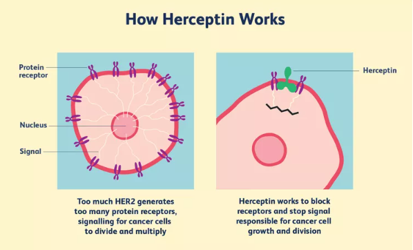
Targeted therapy is a type of cancer treatment that works differently to chemotherapy. Chemotherapy attacks all rapidly dividing cells in the body, but targeted therapy works by “targeting” those differences that help a cancer cell to survive and grow.

Targeted treatments can be taken orally, injected or given intravenously (through a vein). They may be given along with chemotherapy.

Side effects of targeted therapy can include fever, headache, and a rash; the targeted therapy trastuzumab (brand name Herceptin) may also affect heart function.

Cancer treatment centres in Nepal

1.   B. P. Koirala Memorial Cancer Hospital (BPKMCH):

BPKMCH is the first tertiary level cancer hospital in Nepal, which provides all types of cancer treatment services, such as surgical oncology, medical oncology, radiation oncology, pathology services, radio diagnosis imaging and nuclear medicine services.

  • Kathmandu Cancer Centre
  • Bhaktapur Cancer Hospital
  • Nepal Cancer Hospital and Research Centre
  • Suhil Koirala Prakhar Cancer Hospital
  • B & C Medical College Teaching Hospital & Research Center
  • Purbanchal Cancer Hospital
  • Cmc Cancer Institute , Bharatpur , Chitwan

There are also other tertiary care centres for cancer treatment such as Tribhuvan University Teaching Hospital (IOM), Bir Hospital, medical colleges and private cancer centres in Nepal.

Treatment of breast cancer

Treatments are less invasive when breast cancer is detected at an early stage. Challenges for breast cancer treatment in Nepal include patient’s financial status (given the huge out-of-pocket expenditure), accessibility and availability of health services and infrastructures, etc.

Several modalities of treatment are currently available for breast cancer patients as following:

Surgery: Many breast cancer patients go under surgery in order to remove breast cancer. Normally some lymph nodes are removed from under the arm and observed under a microscope to verify if they contain cancerous cells. Surgery to preserve the breast: an operation to remove cancer but not the breast. It includes the following procedures:

  • Lumpectomy: surgery to remove the tumor (mass) and a small quantity of normal tissue around it.
  • Partial mastectomy: surgery to remove a part of the breast that has cancer and some normal tissue surrounding it. The protecting layers over pectoral muscles under the cancer can also be removed. This procedure is also called segmentary mastectomy.

Patients being treated with surgery to preserve the breast can also have some lymph nodes removed from under the arm to perform a biopsy. This procedure is called lymph node dissection and it can be done at the same time than the surgery to preserve the breast or later. The dissection of lymph nodes is done through a separated incision.

Other types of surgery include the following procedures:

  • Total mastectomy: surgery that removes the breast that contains cancer. This procedure is also called simple mastectomy. Some of the lymph nodes can be removed from under the arm to perform a biopsy at the same moment of the surgery or after it. This is done through a separate intersection.
  • Modified radical mastectomy: surgery to remove the breast that contains cancer, many of the lymph nodes under the arm, protecting layers of pectoral muscles and, sometimes, part of the muscles of the breast wall.

Chemotherapy can be administrated before the surgery to remove the tumor. When chemotherapy is administrated before the surgery, it may reduce the size of the tumor and quantity of tissue that needs to be removed during surgery. Treatment administrated before surgery is called neoadjuvant therapy. Even if the doctor removes all the cancer in the surgery, some patients may receive radiotherapy, chemotherapy, or therapy with hormones after the surgery in order to destroy any cancerous cells

that may have been left. The treatment administrated after the surgery to reduce the risk of cancer returning is called adjuvant therapy.