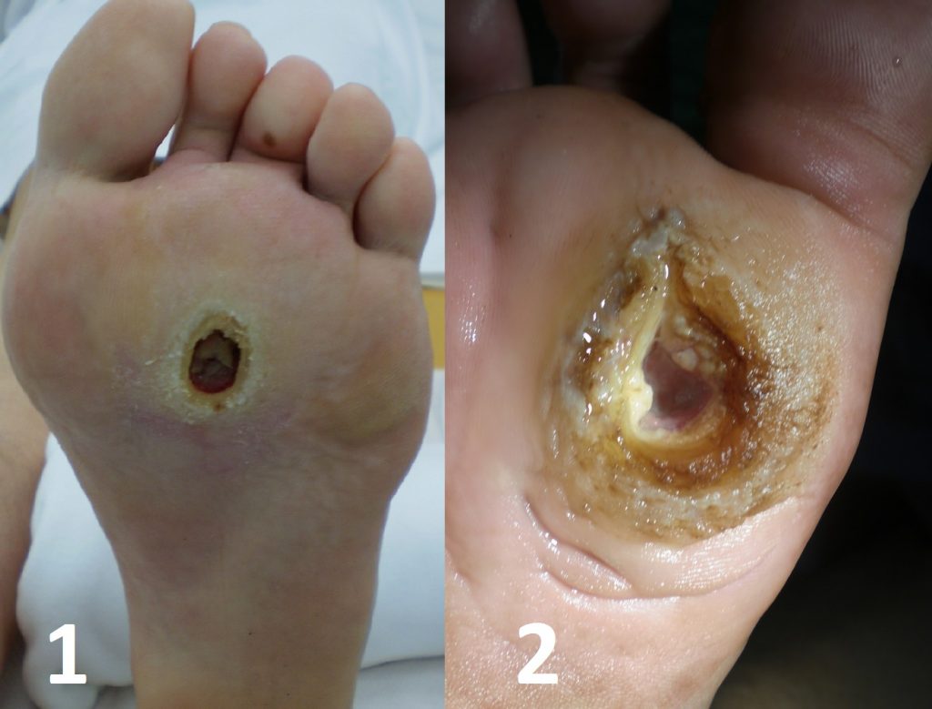
Ulcers are defined as abnormal breaks in the skin or mucous membranes. They can be caused by a wide number of pathologies and have a prevalence of approximately 1%.
The majority of lower limb ulcers have a venous origin (80%), with other common causes including arterial insufficiency and diabetic-related neuropathy. Rarely, they can also be caused by infection, trauma, vasculitis or malignancy (typically squamous cell carcinoma).

Key Points
- Venous ulcers are shallow ulcers with a granulated base, often with other clinical features of venous insufficiency present
- Neuropathic ulcers are painless ulcers over areas of abnormal pressure, often secondary to joint deformity in diabetics
- Arterial ulcers are found at distal sites, often with well-defined borders and other evidence of arterial insufficiency
Risk Factors
The risk factors for developing a venous ulcer include:
- Increasing age
- Pre-existing venous incompetence or history of venous thromboembolism
- Including varicose veins
- Pregnancy
- Obesity or physical inactivity
- Severe leg injury or trauma
Investigations
The diagnosis of venous ulcers is clinical, with the underlying venous insufficiency confirmed by Duplex Ultrasound. Most commonly venous incompetence occurs at the sapheno-femoral or sapheno-popliteal junctions, although it may occur in any perforator.
An Ankle Brachial Pressure Index (ABPI) is required to assess for any arterial component to the ulcers and to determine whether compression therapy will be suitable. If infection is suspected (i.e. erythematous or with purulent exudate) then consider microbiology swabs and antibiotics.
Take swab cultures if suspecting an associated infection. Consider a thrombophilia and vasculitic screening in young patients, especially if there is a suspicion or family history of prothrombotic and autoimmune diseases.
Arterial Ulcers
An arterial ulcer refers to an ulcer caused by a reduction in arterial blood flow, leading to decreased perfusion of the tissues and subsequent poor healing.
They often form as small deep lesions with well-defined borders and a necrotic base. They most commonly occur distally at sites of trauma and in pressure areas (e.g the heel).
Risk Factors
The main risk factors are those of peripheral arterial disease, including smoking, diabetes mellitus, hypertension, hyperlipidaemia, increasing age, positive family history, and obesity and physical inactivity
Clinical Features
A patient with a suspected arterial ulcer is likely to give a preceding history of intermittent claudication (pain when they walk) or critical limb ischaemia (pain at night).
The ulcer may be painful and often develops over a long period of time, with little to no healing (therefore no or little granulation tissue). Other associated signs include cold limbs, thickened nails, necrotic toes and hair loss.
On examination, the limbs will be cold and have reduced or absent pulses. In pure arterial ulcers, sensation is maintained (unlike neuropathic ulcers). Assess for signs of venous insufficiency, as some patients have mixed pathology.
